From Beans to Biology: Our Kidney Dissection
25th October 2025
In our recent biology lesson, we were introduced to one of the most hands-on activities yet—a dissection. The lab tables were set up with trays, scalpels, scissors, and tweezers, all ready for us to begin.
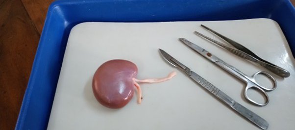
On approaching the tables, I saw something bean-shaped resting in the dissection bath. But it was much bigger than any bean I’d ever seen. It was a goat’s kidney!
As a vegetarian, the sight wasn’t the most appealing. Yet, from an educational perspective, it was an exciting opportunity to witness biology in action.
We began by observing the external structures of the kidney: the tough renal capsule, the ureter, and the blood vessels—the renal artery and vein. Then came the moment of dissection. My teammates carefully sliced the kidney open, revealing a symmetrical structure that looked almost like flower petals hidden inside a bean.
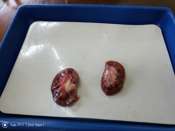
After applying a stain, we were able to clearly view the internal structures: the cortex and its tubules, and the medulla. Seeing it up close was eye-opening.
What struck me most was how different the kidney looked compared to the neat, simplified textbook diagrams we usually study. In real life, the organ was far more intricate and complex.
This dissection brought our biology lesson to life and gave us a stronger foundation for our upcoming topic on the excretory system.
Dhara Shah
Year 11 Student